
About RS Maitri Clinic
Dr.Vaibhav Saxena is trusted and best urologist doctor and Dr. Nandini Ramaswamy is reconstructive and plastic surgeon in Ghaziabad and North India. Visit R S Maitri Urology & Plastic Surgery Clinic now in ghaziabad.
R S Maitri Clinic
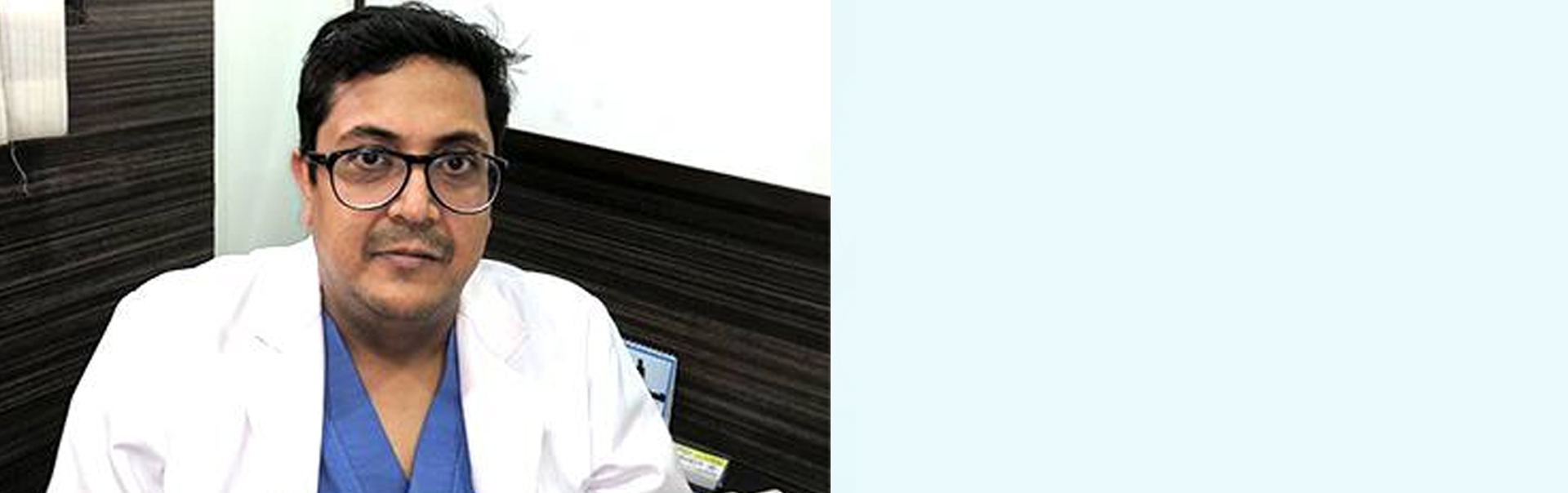
Dr.Vaibhav Saxena
Dr. Nandini Ramaswamy
Emergency Contact
: +91-8527066995
Our Urology Services

Urinary Incontinence
Urinary incontinence is the involuntary leakage of urine. It means a person urinates when they do not want to.

Kidney cancer
Kidney cancer also called renal cancer is a disease in which kidney cells become malignant....

Uro-Oncology
Uro- Oncology offers compassionate care for those diagnosed with kidney cancer, bladder and testicular cancer.

Prostate Cancer
Prostate cancer is one of the most common types of cancer in men. Usually that develops in the prostate gland.....

Urinary calculi
Urinary calculi are solid particles like stones in the urinary system. They can develop anywhere along....

Laparoscopic Surgery
Laparoscopic Surgery has been called one of the greatest advances in the war on cancer and many experts....
Aesthtic & Reconstructive Surgery Services

Nose Reshaping
Nose is the most prominent facial feature which can either make your appearance attractive or ugly

Face-Lift
Facial aging results in a loss of skin elasticity and firmness, sagging tissue and wrinkles........

Liposuction
Technique to remove fat from specific areas of the body, such as the abdomen, hips, thighs, buttocks, arms or neck.

Double Chin
procedure removes fat from beneath the skin and sculpts the chin and neck contour.......

Breast Surgery
There are many different reasons why women seek help including cosmetic concerns, cancer treatment and changes in shape

Tummy Tuck
A tummy tuck, also known as abdominoplasty, removes excess fat and skin and restores.......